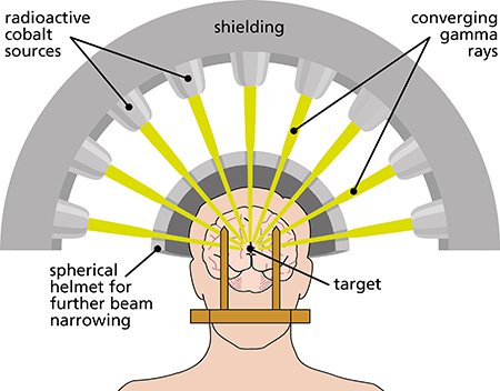
Stereotactic neurosurgery entails the mapping of the brain within a three-dimensional coordinate system, utilizing MRI and CT scans alongside 3D computer workstations. Neurosurgeons can precisely target specific areas of the brain by referencing their 3D coordinates. Stereotactic neurosurgery is generally categorized into stereotactic biopsy and stereotactic brain surgery.
"Stereotactic biopsy" is a surgical procedure in which a neurosurgeon uses a thin needle to extract a small piece of brain tissue for microscopic examination. The purpose of this procedure is to diagnose abnormalities identified on an MRI or CT scan. While these imaging techniques effectively reveal abnormal brain regions, they cannot definitively determine the nature of the abnormality—whether it is a cancerous tumor, a benign tumor, an infection, an inflammatory process, a vascular abnormality, or another pathology. Given the distinct treatment approaches for these conditions, accurately identifying the nature of the lesion is crucial to recommend appropriate treatment.
Stereotactic procedures can be performed either with a "frame" or "frameless," depending on the surgeon's preference and the characteristics of the lesion's location and size.
In the case of a frameless biopsy, a special MRI or CT scan is obtained one or two days before the scheduled biopsy. This image is imported into a computer system, generating a 3-dimensional image of the brain and the biopsy target during the surgery. Utilizing this image alongside a biopsy guidance arm, the surgeon navigates the needle safely to the tumor target.
For a frame-based biopsy, a device called a stereotactic frame is affixed to the patient's head before obtaining the MRI or CT scan to be utilized with the image guidance system. The frame proves particularly beneficial for directing the biopsy needle to small or deeper targets within the brain. Both approaches boast comparable safety and success records. In both frame and frameless biopsies, a small incision is made, and a small hole is drilled in the skull to allow the needle access to the target. At the conclusion of the operation, the needle is extracted, and the incision is closed with sutures or staples. The procedure can be conducted under general anesthesia or with sedation, depending on whether the patient is asleep or drowsy but not fully unconscious.
"Stereotactic brain surgery" is a surgical procedure in which a lesion, often a brain tumor, is removed with the aid of image guidance. Pre-existing images, typically an MRI, are utilized to guide the surgeon precisely to the location of the lesion. This facilitates the creation of an accurate pathway through the brain, ensuring the safe removal of as much abnormal tissue as possible while preserving the integrity of normal, healthy brain tissue.
The primary benefit of stereotactic procedures lies in the millimetric precision achievable, allowing for the precise targeting of abnormalities, even those situated deep inside the brain, using a thin needle. The procedure can be conducted under general anesthesia or with sedation. Patients undergo monitoring for several hours post-procedure and typically return home within a day.
Goutam Neuro Care boasts a cutting-edge CRW stereotactic system, complemented by the advanced capabilities of the 3.0 Tesla MRI and Multislice CT systems. This combination ensures the highest level of precision during stereotactic procedures.
 Stereotactic Surgery
© Goutam Neuro Care. All Rights Reserviced. Developed & Digital Marketing by DGTAL HEALTHCARE MARKETING AGENCY